
Vet’s diary: a fresh challenge
This time, Peterborough vet Laura Frost shares a challenging yet satisfying case of a kind she had never had to deal with before
After eight years in practice I have experience with most emergency situations. I am confident with twisted stomachs, ruptured diaphragms, bleeding and breaks. However, one thing I had never come across was a pneumothorax (punctured lung). A pneumothorax can develop for several reasons and can be classed as open or closed. An open pneumothorax is usually trauma related. A penetrating injury to the chest wall leaves the space around the lungs connected to the outside world. The loss of normal pressure inside the chest causes air from the atmosphere to be sucked into the chest which then compresses the lung. A closed pneumothorax results from rupture of lung tissue causing air to leak directly from the lung into the chest cavity. An open pneumothorax is usually an easy diagnosis because there is an obvious wound to the chest. A closed pneumothorax however is much trickier to spot.
This dog was a young Lurcher who had run free from his owner and gone missing for several hours. He came back covered in small scratches and ‘out of breath’. His owner took him home to bathe his wounds and let him recover from the afternoon’s excitement only to find his breathing did not return to normal and his chest appeared swollen to one side. Understandably worried, they rushed him in to the surgery wondering if he had been hit by a car. On examination the left side of the chest was painful and bruised. His breathing was rapid but he still walked around the room sniffing the floor and wagging his tail.
At first I thought the increased breathing might be pain related, so I gave some injections and monitored him. No improvement was seen. Although I considered a pneumothorax possible, I always expected the breathing difficulty to be worse than this. The dog looked relatively happy and alert. I asked the owner about taking a chest x-ray, just to be on the safe side. And am I glad I took that x-ray! Air is usually black on x-rays and soft tissue is white, with bone being a really bright white. The lungs normally appear as a white pattern indicating the blood vessels and connective tissue with black air laced in between. This x-ray was definitely not normal. The entire left lung was squashed to a quarter of its original size showing a dense white and airless structure surrounded by a large expanse of black air between the lung and the chest wall.
I was surprised by how well the dog was with such a dramatic reduction in lung volume. He was still wagging his tail and his gums were still nice and pink. Animals really are amazing. I am sorry to dispel a myth (for all the Holby City fans out there), but there was no sudden dash across the room to grab a needle to dramatically stab into the chest followed by a whoosh of air that pulled the patient back from the brink. There was plenty of time for a calmer, more measured approach.
We started by clipping a patch of hair from the side of his chest and a small amount of local anaesthetic was injected into the skin and muscle. While the local worked its magic I connected a butterfly needle to a three-way tap and a 20ml syringe. The three-way tap allows air to be removed from the chest into the syringe, then as the tap is turned it is squirted into the atmosphere leaving the syringe ready to use again. This technique is really important because it ensures that no further air from the room is sucked into the chest. It also allows us to measure the volume of air removed.
Two thousand, four hundred millilitres later, the chest was drained of air. The dog’s breathing rate slowed progressively and returned to normal. The next stepping stone was whether any more air was going to continue to leak from the damaged lung. In most trauma cases the damage can seal itself over quite quickly, but occasionally surgery is needed. Unfortunately by next morning his breathing had deteriorated again. A further x-ray showed that the chest had again filled with air. We repeated the drainage procedure but only 1000ml was removed this time. Rather than rushing to surgery I decided to monitor for the day and later repeated the drainage. This time only 600ml was removed. The leak was slowing down. One more night of care and the danger passed. He went home with strict instructions to rest and have some TLC from his loving owner – and I am glad to report that he has since made a full recovery.
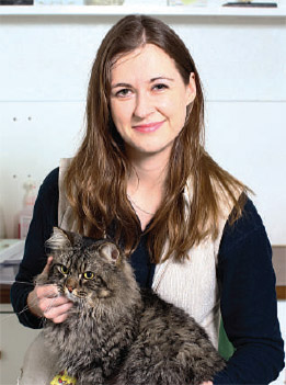 Laura Frost MRCVS MA VetMB is a vet at local veterinary practice Pengelly and Mizen.
Pengelly and Mizen Veterinary Surgeons 89-93 Park Road, Peterborough PE1 2TR T: 01733 554953 Emergencies only: 01733 896000 www.pengellyandmizen.co.uk
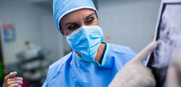
Image: www.freepik.com/free-photo/surgeons-discussing-on-patient-x-ray-in-operation-room_1008454